 +91 96001 07057
 Sidharam Heart Clinic Adyar, Gandhi Nagar, Canal Bank Road, Opp.St.Louis School, Adyar, Chennai, Tamil Nadu 600020
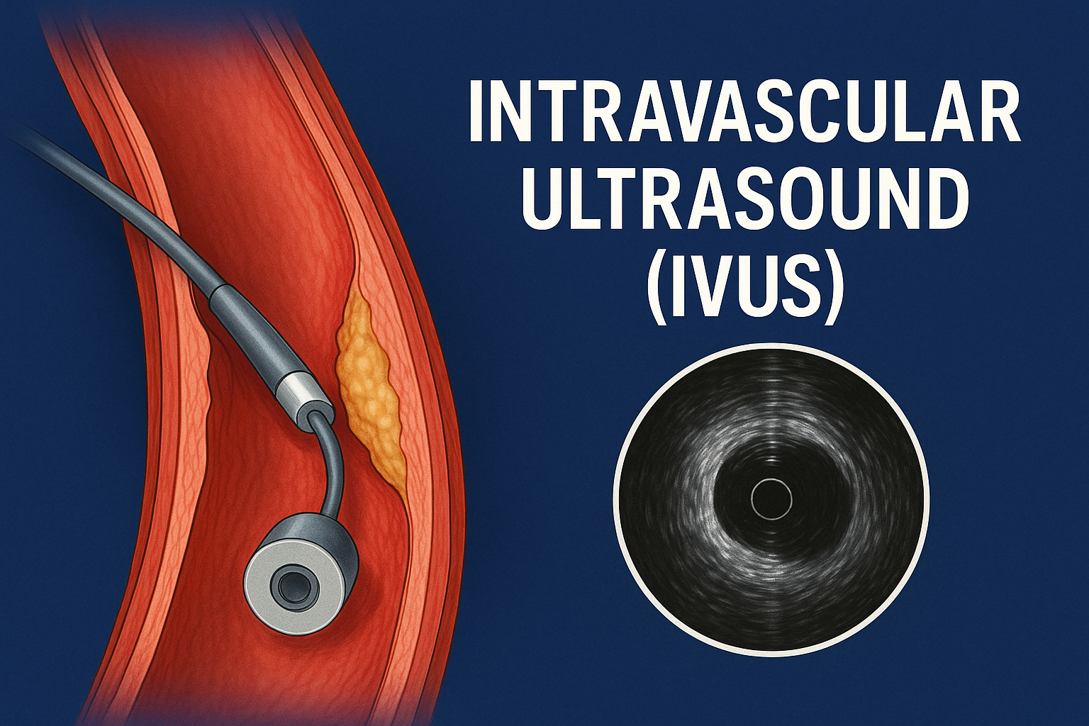 13 May, 2025
A Closer Look Inside the Arteries: How IVUS is Revolutionising Heart Care
When it comes to treating heart disease, precision is everything. And for Dr. Dhamodaran—one of the most respected interventional cardiologists in India—precision starts with clarity. That’s where IVUS steps in.
Short for Intravascular Ultrasound, IVUS is not your typical heart scan. While traditional angiograms sketch the roadmap of your arteries, IVUS dives beneath the surface—right into the layers of the arterial wall. For patients battling coronary artery disease (CAD), that difference can be life-changing.
In this guide, we’ll explore what IVUS is, how it works, and why Dr. Dhamodaran uses this advanced imaging technique to ensure no detail is left to guesswork.
Meet the Doctor Behind the Screen
Dr. Dhamodaran is among India’s top-ranked interventional cardiologists, known for his deep expertise in complex cardiac procedures and a commitment to patient-first care. With extensive training and decades of experience, he brings the best of modern cardiac science to the heart of Chennai.
Whether you’re a first-time patient or managing chronic heart disease, you’re in expert hands.
What is IVUS?
IVUS (Intravascular Ultrasound) is a catheter-based imaging method that uses sound waves to capture real-time, high-resolution images from inside your blood vessels. It provides a 360-degree view of the coronary artery wall, allowing doctors to measure plaque, assess blockages, and guide treatment.
Instead of only looking at the blood flow channel (lumen), IVUS reveals what’s happening within the walls of the artery—an essential feature when decisions need to be made during stent placement or angioplasty.
Why Is IVUS Better Than a Regular Angiogram?
While angiography has long been a trusted diagnostic tool, it comes with its limits. It shows where a narrowing is but doesn’t always reveal why or how bad it is.
Here’s how IVUS fills the gap:
| Aspect | Angiography | IVUS |
|---|---|---|
| Views the artery’s interior | ✅ | ✅ |
| Shows plaque behind vessel wall | ❌ | ✅ |
| Helps guide stent size and placement | ⚠️ Estimations only | ✅ Accurate measurements |
| Distinguishes plaque types (hard vs. soft) | ❌ | ✅ |
| Identifies hidden lesions | ❌ | ✅ |
Bottom line? IVUS doesn’t just show where the problem is—it shows what the problem is made of.
How IVUS Works: From Insertion to Insight
IVUS is performed during a cardiac catheterisation. Here’s how it happens:
-
Access Point: A thin, flexible catheter is inserted into the artery, usually via the wrist (radial) or groin (femoral).
-
Ultrasound Probe: At the catheter’s tip is a mini ultrasound transducer that emits sound waves.
-
Image Capture: These waves reflect off vessel walls, creating a layered image of the artery.
-
On-Screen Analysis: The real-time scan appears on a monitor, giving the cardiologist a 360° cross-section of the vessel.
When is IVUS Recommended?
Dr. Dhamodaran may recommend IVUS in various scenarios, particularly when angiography alone is insufficient. Common situations include:
🔍 Ambiguous or Borderline Lesions
Sometimes, a narrowing might look mild on angiography—but IVUS may uncover a severe plaque burden hidden in the vessel wall.
🫀 Left Main Artery Assessment
The left main coronary artery is critical. Misjudging its blockage could be fatal. IVUS offers accurate measurements to confirm whether to stent or not.
🧱 Complex or Calcified Plaques
Some blockages are made of hard calcium. IVUS helps characterise the plaque and guide whether rotational atherectomy or balloon preparation is needed before stenting.
🧩 Stent Optimisation
IVUS ensures the stent is fully opened and pressed against the artery walls, reducing the risk of complications like restenosis or thrombosis.
Real-World Case: How IVUS Can Change Outcomes
Let’s take an example:
Patient: 60-year-old male with diabetes and chest tightness.
Initial Angiogram: Shows 50% blockage in the left main artery. Not clear if intervention is needed.
IVUS Result:
-
Lumen area = 4.0 mm² (normal >6 mm²)
-
Heavy plaque burden with early calcification
Decision: Stent required. IVUS helps pick the right stent size and ensures it’s properly deployed.
Outcome: Better perfusion, reduced chest pain, and lower risk of future cardiac events.
Advantages of Using IVUS
IVUS isn’t just a cool tech toy—it offers life-saving advantages:
-
✅ Precision: Identifies plaque not visible on angiography
-
✅ Personalised Treatment: Selects accurate stent size based on actual vessel dimensions
-
✅ Reduced Risk: Ensures optimal stent expansion and apposition
-
✅ Improved Outcomes: Leads to fewer complications and repeat procedures
-
✅ Deeper Insight: Helps assess vessel healing in follow-ups
Are There Any Limitations?
While IVUS is a powerful tool, it has its constraints:
-
❌ Slightly higher cost compared to angiography alone
-
❌ Requires specialist expertise to interpret accurately
-
❌ Adds a few minutes to the procedure time
-
❌ Not always available in all centres
However, in expert hands like Dr. Dhamodaran’s, the benefits far outweigh the limitations—especially for patients with complex cardiac profiles.
IVUS vs OCT vs Angiography
Here’s a quick comparison to help understand how IVUS stacks up against other tools:
| Feature | IVUS | OCT (Optical Coherence Tomography) | Angiography |
|---|---|---|---|
| Imaging Method | Ultrasound | Light | X-ray with dye |
| Resolution | Moderate | Very High | Low |
| Depth Penetration | Deep | Superficial | Surface Only |
| Ideal Use | Plaque assessment, stent sizing | Stent detail, fine structures | Lumen shape and flow |
| Blood Clearance Needed | No | Yes | No |
What Patients Should Know Before an IVUS Procedure
💡 Will I be awake?
Yes, IVUS is usually done during angioplasty or a diagnostic catheterisation, under local anaesthesia and mild sedation.
💡 Will I feel the catheter?
No. Patients don’t feel the IVUS catheter. The entire process is smooth and pain-free.
💡 Does it increase risk?
Not when done by trained professionals. In fact, it reduces long-term risk by ensuring precise treatment.
Frequently Asked Questions (FAQs)
1. What does IVUS help diagnose?
It helps identify the amount, type, and extent of plaque inside your coronary arteries. It also aids in deciding if a stent is needed.
2. Is IVUS used in all heart procedures?
No. It’s reserved for complex or unclear cases where additional imaging is necessary for decision-making.
3. How long does it take?
It adds about 5–10 minutes to a typical procedure.
4. Can IVUS be done without stenting?
Yes. It’s often used just for diagnostic clarity, especially when angiographic findings are ambiguous.
5. Does insurance cover IVUS?
In many hospital setups, IVUS is included under the broader catheterisation or angioplasty coverage. Check with your provider for specifics.
6. Is IVUS safe for older patients?
Yes. It’s safe for all age groups, especially when performed by experienced cardiologists like Dr. Dhamodaran.
💬 Consult Dr. Dhamodaran
Dr. Dhamodaran
Renowned Interventional Cardiologist, Expert in Complex PCI & Imaging-Guided Interventions
🏥 Sidharam Multispeciality Clinic
📍 Old #2, New #4, Canal Bank Road, Gandhi Nagar, Adyar, Chennai, Tamil Nadu 600020
🕒 MON – THU: 6 PM to 9 PM
🏥 Apollo 247, Apollo Hospitals
📍 Greams Road, Chennai
🕒 MON – SAT: 10 AM to 4 PM