 +91 96001 07057
 Sidharam Heart Clinic Adyar, Gandhi Nagar, Canal Bank Road, Opp.St.Louis School, Adyar, Chennai, Tamil Nadu 600020
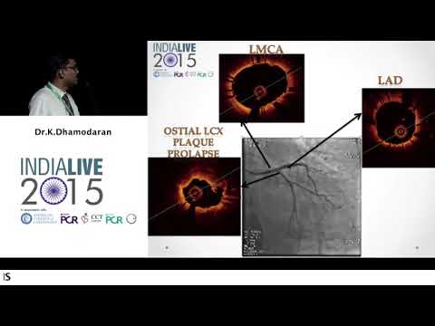 9 February, 2025
Intra Procedural Stent Thrombosis: A Case Study
In this detailed case study, we explore the complexities of intra procedural stent thrombosis in a 65-year-old male patient. Through a step-by-step analysis of the case, we will highlight key technical strategies, potential complications, and important learning points for interventional cardiologists.
Introduction to the Case
This case revolves around a 65-year-old male patient, a known smoker with a history of hypertension but no diabetes. The patient presented with an evolved inferior myocardial infarction and ongoing angina symptoms. The urgency of the situation was underscored by the patient’s left ventricular dysfunction, prompting immediate intervention.
Understanding the patient’s profile is vital. His cardiovascular risk factors, coupled with the acute presentation, set the stage for a complex intervention. This case highlights the importance of meticulous planning in interventional cardiology, particularly when dealing with potential complications during procedures.
Patient Profile and Presentation
The patient, a 65-year-old male, had several significant risk factors, including smoking and hypertension. Upon presentation, he exhibited symptoms consistent with an inferior myocardial infarction. Notably, his left ventricular function was compromised, which necessitated a careful approach to his treatment.
During the evaluation, there were no signs of significant mitral regurgitation or pulmonary hypertension, which is essential in assessing the patient’s overall cardiac status. The coronary angiogram revealed important details about the patient’s vascular condition, particularly affecting the left circumflex artery (LCX) and left anterior descending artery (LAD).
Coronary Angiogram
Coronary Angiogram
Findings
The coronary angiogram displayed critical insights into the patient’s condition. The LCX and LAD showed minor blockages, particularly at the osteoproximal segment. The bifurcation angle between the LAD and LCX was approximately 70 to 80 degrees, which is noteworthy for planning stent deployment.
With the potential for plaque instability, particularly in the LCX, the angiogram informed the initial approach. It was crucial to consider the risk of plaque shifting during balloon dilation, which could complicate the procedure. The findings necessitated a proactive strategy to ensure vessel integrity throughout the intervention.
Initial Treatment Plan
The initial treatment plan focused on protecting the LAD while addressing the LCX. A wire protection strategy was employed, particularly because the LAD showed no significant occlusion at the outset. The concern was that any disturbance in the LCX could lead to complications in the LAD during the intervention.
This strategy included pre-dilating the LCX with a 2.5 x 15 mm balloon. The aim was to establish a pathway for stent deployment while minimizing the risk of plaque movement. A backup plan was also devised, allowing for a crossover intervention if necessary.
Deployment of Stents
Following the pre-dilation, a 3 x 15 mm drug-eluting stent was deployed in the LCX. Careful attention was paid to the positioning of the stent to avoid migration or excessive protrusion into the left main artery. Despite meticulous planning, a nearly 90% stenosis was observed in the LAD post-angioplasty, even though the patient remained asymptomatic.
Given the ECG changes, it was evident that a shift in strategy was necessary. The decision was made to proceed with a crossover from the left main to LAD, utilizing a 3.5 x 23 mm stent. This adjustment highlighted the dynamic nature of interventional procedures and the need for real-time decision-making based on the patient’s response.
Post-Dilation and Kissing Balloon Dilatation
Post-dilation of the left main artery was performed to enhance stent apposition and ensure optimal flow dynamics. This was followed by kissing balloon dilatation, which is critical in bifurcation lesions to maintain patency in both branches. The results appeared satisfactory, with no significant edge dissection or thrombus formation noted immediately after the procedure.
However, despite these positive outcomes, the patient experienced acute hemodynamic instability shortly after the intervention. This underscored the unpredictable nature of such procedures and the necessity for rapid assessment and intervention in the event of complications.
Patient’s Deterioration and Emergencies
Shortly after the procedure, the patient’s condition took a sudden turn. Within ten minutes, he developed severe bradycardia and a rapid decline in blood pressure, raising immediate concerns. The swift deterioration prompted a reassessment of the situation, leading to a suspicion of left main occlusion.
In such emergencies, timely intervention is crucial. A repeat angiogram revealed a total occlusion of the LCX, while the LED exhibited slow flow. This unexpected development highlighted the need for a deeper understanding of the underlying causes, which were suspected to be linked to hypertension rather than direct embolization from the stent.
Rewiring and Aspirating the LCX
Addressing the total occlusion required a meticulous approach. The first step involved rewiring the LCX through multiple stent layers, akin to a modified tree stenting technique. This process is particularly challenging due to the acute angles and non-compliant nature of the vessel.
After successfully navigating the wire, the next phase involved using an aspiration catheter to remove any thrombus that may have contributed to the occlusion. The aspiration technique was selected over thrombectomy due to the low thrombus burden observed, allowing for a prolonged balloon inflation strategy instead.
This decision was pivotal, as using a bulky catheter in a constrained environment can be technically demanding. However, the focus remained on achieving a stable flow in the LED while ensuring the integrity of the LCX.
Final Angiogram and OCT Analysis
Following the interventions, a final angiogram was conducted to assess the outcomes. The results indicated a significant improvement, yet there were lingering concerns regarding the haziness at the inner border of the LCX ostium. This prompted further investigation using Optical Coherence Tomography (OCT), although it was not available immediately following the procedure.
Four days later, a follow-up angiogram was performed, revealing some retraction of the stent and narrowing at the ostium, likely due to the acute angle. The OCT analysis later provided insights into the mechanisms behind the stent thrombosis, revealing tissue prolapse that could be attributed to either plaque collapse or thrombus prolapse.
Understanding these dynamics is essential. The OCT images showed irregular borders, indicating potential complications that could affect long-term outcomes. A thorough analysis of tissue attenuation was necessary to differentiate between thrombus-related issues and structural vessel collapse.
Learning Points from the Case
This case underscores several critical learning points for interventional cardiologists. First and foremost, meticulous pre-procedural planning is imperative. Even in seemingly straightforward cases, unexpected complications can arise, necessitating contingency plans for managing potential vessel compromise.
Moreover, the importance of wire protection cannot be overstated. Implementing wire protection strategies, particularly in bifurcation lesions, can prevent plaque shift and safeguard the integrity of adjacent vessels.
Additionally, this case illustrates the necessity for rapid assessment and intervention during emergencies. Being prepared to adapt strategies, such as transitioning from stenting to balloon inflation or utilizing thrombus aspiration techniques, can significantly impact patient outcomes.
Review of Literature and Key Trials
A review of the literature reveals valuable insights into intra procedural stent thrombosis (IPST). Notably, the Champion Phoenix trial highlighted the significance of IPST as a predictor of both short-term and long-term mortality. The study emphasized that IPST is not merely a procedural complication but a critical factor influencing patient prognosis.
Several factors contribute to IPST, including patient characteristics such as diabetes, lesion complexities like ruptured plaques, and technical aspects such as stent placement and balloon inflation pressures. Understanding these variables is crucial for developing effective strategies to mitigate risks during procedures.
Furthermore, the literature stresses the importance of imaging modalities in identifying the mechanisms behind complications. While OCT provides detailed insights, the availability of such technology in cath labs is essential for timely decision-making and patient management.
Conclusion and Recommendations
In conclusion, this case serves as a poignant reminder of the complexities inherent in interventional cardiology. It emphasizes the need for thorough pre-procedural planning, the importance of wire protection, and the necessity for rapid adaptation in response to unforeseen complications.
Recommendations for practice include maintaining a high index of suspicion for complications, particularly in patients with significant cardiovascular risk factors. Additionally, interventional cardiologists should ensure access to advanced imaging modalities to facilitate better decision-making during complex procedures.
Ultimately, this case illustrates that even with meticulous planning, the unpredictable nature of cardiovascular interventions necessitates a flexible approach and a commitment to continuous learning and improvement.