 +91 96001 07057
 Sidharam Heart Clinic Adyar, Gandhi Nagar, Canal Bank Road, Opp.St.Louis School, Adyar, Chennai, Tamil Nadu 600020
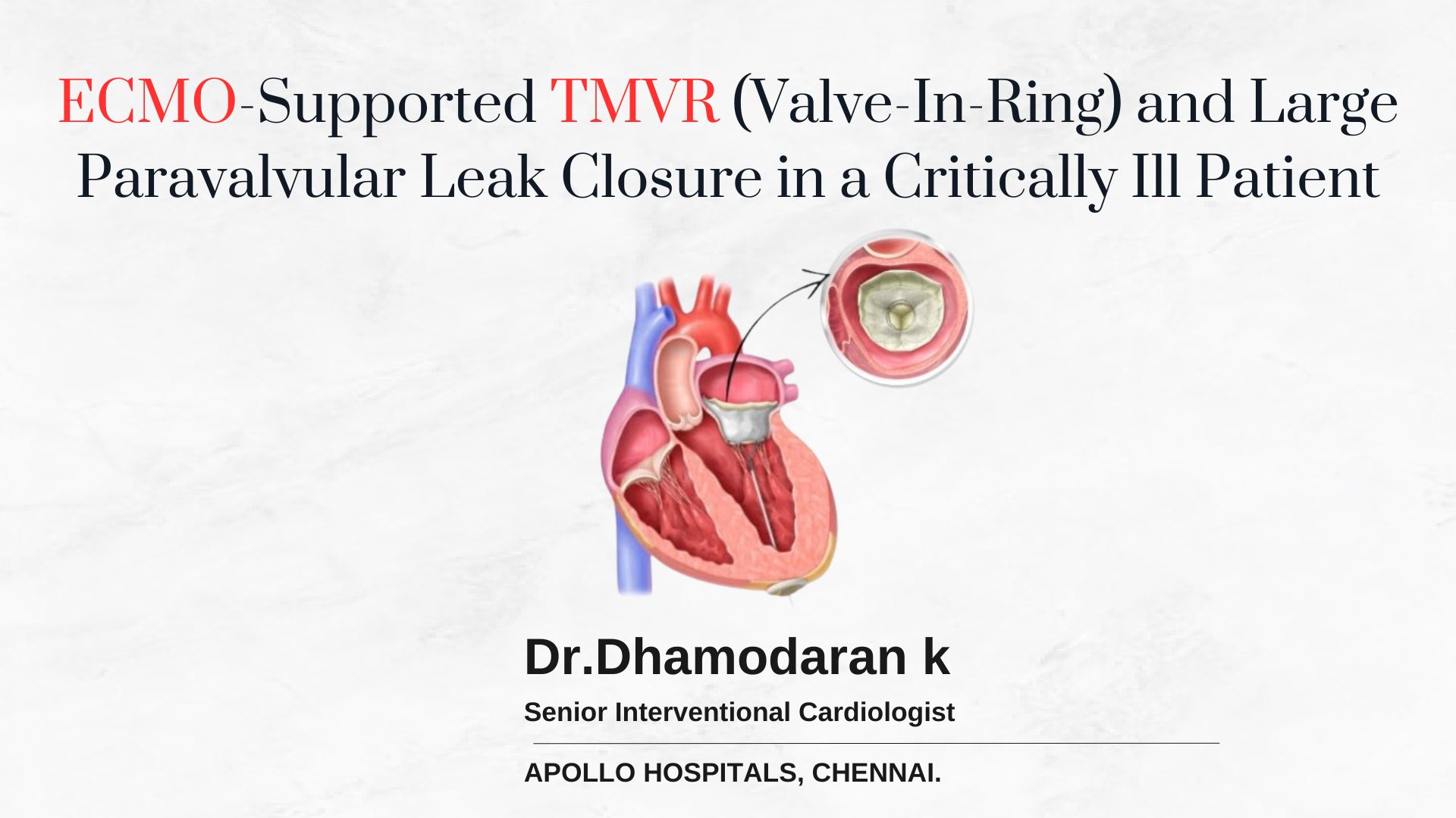 3 January, 2025
Sharing a Case of ECMO-Supported TMVR (Valve in Ring) with Para-Valvular Leak Closure in a Critically Ill Patient
Patient Background
A 56-year-old male with a history of chronic smoking, chronic kidney disease (CKD), and coronary artery disease (CAD) presented with acute decompensated heart failure (ADHF). He had previously suffered an inferior wall myocardial infarction (IWMI) and mild left ventricular (LV) dysfunction managed conservatively. Additionally, he was diagnosed with antiphospholipid antibody syndrome, which had led to axillary artery and cortical vein thrombosis.
Clinical History:
- 1.5 years ago, the patient underwent coronary artery bypass grafting (CABG) and mitral valve (MV) repair with a 28 mm SJM Rigid Saddle Ring for severe mitral regurgitation (MR) caused by chordal rupture. Post-surgery, he experienced symptomatic improvement for three months before being hospitalized again for acute heart failure, severe MR, pulmonary arterial hypertension (PAH), and worsening renal function.
- The patient was managed conservatively for heart failure and maintained on hemodialysis until his transfer to our facility one month prior to the current admission.
Admission Presentation
On admission, the patient presented with the following:
- Symptoms: Severe shortness of breath (ambulatory Class IV), orthopnea, and anasarca.
- Vitals: Blood pressure: 90/60 mmHg; Hemoglobin: 8%; Urea: 104 mg/dL; Serum creatinine: 4 mg/dL; Raised liver function tests (LFTs), despite maintenance hemodialysis.
- Imaging:
- TTE and TEE confirmed severe intravalvular and paravalvular leaks (25×12 mm).
- LVEF: 48%; RVSP: 80 mmHg; RVFAC: 18%.
Heart Team Evaluation
Given the patient’s critically ill status and multiple comorbidities, a multidisciplinary heart team decided on transcatheter mitral valve replacement (TMVR) with para-valvular leak (PVL) closure as a single-stage procedure under hemodynamic support.
- STS and EuroSCORE: Prohibitive risk for surgical mitral valve replacement (SMVR) due to severe PAH, RV dysfunction, and inability to tolerate cardiopulmonary bypass (CPB).
- Challenges Identified:
- Adverse hemodynamics.
- Rigid Saddle Ring posing risks of valve dislodgement and post-THV PVL.
- Neo-LVOT area >350 mm² minimizing the risk of LVOT obstruction.
- Large PVL requiring two AVP II plugs for closure.
Procedural Strategy
- Prophylactic ECMO Placement:
- Ultrasound-guided percutaneous ECMO was initiated with left femoral artery (LFA, 19F) and left femoral vein (LFV, 21F) cannulation, maintaining a 2L flow rate.
- Ultrasound-guided percutaneous ECMO was initiated with left femoral artery (LFA, 19F) and left femoral vein (LFV, 21F) cannulation, maintaining a 2L flow rate.
- Transseptal Puncture (TSP):
- TEE-guided posteroinferior puncture followed by slow valve inflation (20-30 seconds) under rapid pacing.
- TMVR Deployment:
- A 27.5 mm Myval THV was deployed within the rigid saddle ring in a 70:30 position.
- PVL Closure:
- TEE-guided wiring for simultaneous deployment of two AVP II plugs.
- Challenges included one wire slipping out, prolonging procedural time.
- ECMO Decannulation:
- On-table decannulation with Perclose for primary access and ECMO.
Post-Procedural Management
- Immediate Post-Procedure:
- Received 4 units of blood transfusion.
- Inotropes and ventilation continued for 12 hours.
- Day 1 Labs:
- Hemoglobin: 7.5 g/dL; Platelets: 70K; Leukocytosis.
- Elevated LFTs, LDH, and INR (2.1).
- Hematologist consultation revealed DIC likely secondary to sepsis or consumption coagulopathy.
- Recovered over one week except for persistent INR elevation.
Pre-Discharge Evaluation
- Clinical Status: Symptomatically improved with Class II dyspnea, no orthopnea or edema.
- Echocardiography Findings:
- No valvular MR; mild residual PVL.
- LVEF: 60%; RVSP: 50 mmHg; RVFAC: 30%.
- Discharge Plan:
- Single antiplatelet therapy (SAPT).
Follow-Up
- Three Weeks Post-Discharge:
- Symptomatically stable and better.
Summary of Key Outcomes
- Successful TMVR within a rigid saddle ring with concurrent PVL closure.
- Effective use of prophylactic ECMO to maintain hemodynamic stability during the procedure.
- Post-procedure recovery demonstrated marked improvement in functional status and echocardiographic parameters.