 +91 96001 07057
 Sidharam Heart Clinic Adyar, Gandhi Nagar, Canal Bank Road, Opp.St.Louis School, Adyar, Chennai, Tamil Nadu 600020

 17 September, 2026
My Heart Blockage Is Heavily Calcified — Can Angioplasty Still Work?
“You have a heart blockage, but there is a lot of calcium in the artery.”
Patients often hear this after a coronary angiogram and wonder:
“Why does calcium matter? Can’t the doctor simply put in a stent?”
Sometimes it is not that simple.
A heavily calcified coronary artery can behave very differently from a normal artery. The blockage may be extremely hard, balloons may not expand adequately, and a stent may not open completely unless the calcium is first treated appropriately.
The good news is that modern complex angioplasty offers several techniques specifically designed to deal with difficult calcified coronary blockages.
But the correct treatment depends on how much calcium is present, where it is located and how deeply it surrounds the artery.
What Does “Calcium in the Heart Artery” Actually Mean?
Coronary artery disease develops when plaque accumulates within the walls of the arteries supplying the heart.
Over time, some plaques become hardened by deposits of calcium.
Think of the difference between:
A soft blockage that can be compressed relatively easily,
and
a hard shell-like blockage that does not readily move when a balloon is inflated.
That hard calcium is one of the reasons some angioplasty procedures become significantly more challenging.
The Society for Cardiovascular Angiography and Interventions notes that calcified coronary lesions are associated with greater procedural complexity and higher short- and long-term risks compared with non-calcified lesions.
Why Is Calcium a Problem During Angioplasty?
Imagine trying to expand a balloon inside a rigid pipe.
If the artery wall cannot stretch because it is heavily calcified, several problems can occur.
The balloon may not expand
A standard angioplasty balloon may inflate but still fail to adequately open the hardest portion of the blockage.
The stent may not open completely
A coronary stent needs to expand firmly against the artery wall.
If severe calcium prevents complete expansion, parts of the stent may remain underexpanded.
This is important because optimal stent expansion is one of the key goals of PCI.
The artery may be more difficult to navigate
Heavy calcium can make it challenging to deliver balloons, imaging catheters and stents through the diseased segment.
The procedure may become more complex
Calcified lesions may require additional equipment, imaging and specialised techniques before a stent is implanted.
This process is commonly called lesion preparation or calcium modification.
So How Do Doctors Treat a “Rock-Hard” Heart Blockage?
There is no single technique for every calcified artery.
The cardiologist first needs to understand what type of calcium is present.
Depending on the anatomy, several approaches may be considered.
Option 1: Specialised High-Pressure or Scoring Balloons
Some calcified lesions can be prepared with specialised balloons.
These may include:
- Non-compliant balloons
- High-pressure balloons
- Scoring balloons
- Cutting balloons
These devices can sometimes create controlled fractures or modify resistant plaque so the artery can expand more effectively.
However, very severe or deeply circumferential calcium may require a different approach.
Option 2: Rotational Atherectomy
Some patients hear this described as:
“Rotablation.”
Rotational atherectomy uses a very small rotating burr that travels through the calcified segment.
Its role is not simply to “drill out the whole blockage.”
Instead, it modifies particularly resistant calcium and helps create a pathway so that balloons and stents can be delivered and expanded more effectively.
For selected severely calcified lesions, atherectomy remains one of the established tools available to interventional cardiologists.
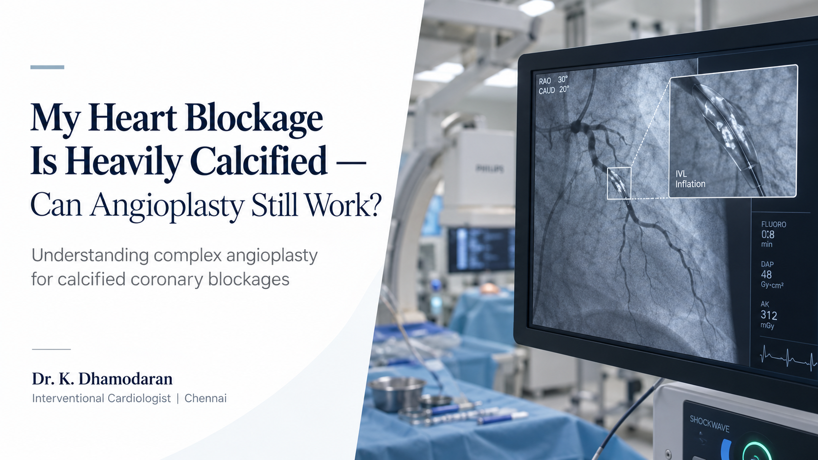
Option 3: Intravascular Lithotripsy — IVL
One of the technologies patients increasingly hear about is:
Intravascular Lithotripsy, or IVL
The principle is similar to the use of shock waves for breaking kidney stones, although coronary IVL is specifically designed for use inside the heart arteries.
A specialised balloon is positioned within the calcified blockage.
The device then delivers controlled acoustic pressure waves that can fracture calcium within the vessel wall.
After the calcium has been modified, the artery may be more capable of expanding with a balloon and accommodating a properly expanded stent.
IVL has become an important option among the calcium-modification technologies available for selected calcified coronary lesions.
Does Every Calcified Blockage Need IVL or Rotablation?
No.
This is important.
The treatment should not be selected simply because calcium appears on an angiogram.
One patient may need only a specialised balloon.
Another may benefit from rotational atherectomy.
Another may be suitable for IVL.
And particularly challenging lesions may sometimes require more than one technique.
The choice depends on factors such as:
How thick the calcium is
How much of the artery circumference it involves
How long the calcified segment is
Whether a balloon can cross the lesion
Whether the balloon can expand
Whether previous stents are present
The diameter and location of the artery
SCAI guidance therefore recommends a tailored approach to calcium modification rather than one treatment for every calcified lesion.
“My Angiogram Already Shows Calcium. Why Do I Need IVUS or OCT?”
This is where modern complex angioplasty becomes particularly interesting.
A conventional coronary angiogram shows the outline of the artery.
But calcium exists inside the wall of the vessel.
That means angiography may not always reveal exactly:
- How thick the calcium is
- How deeply it extends
- Whether it surrounds the artery
- Whether a stent has fully expanded
Two technologies can provide much more detailed information from inside the coronary artery.
IVUS — Intravascular Ultrasound
A tiny ultrasound probe travels through the artery and produces cross-sectional images of the vessel.
OCT — Optical Coherence Tomography
OCT uses light to create very high-resolution images of the artery from inside.
Both can help identify the distribution of calcium and guide the treatment strategy.
SCAI recommends intravascular imaging whenever feasible in calcified coronary disease to help determine calcium-modification strategy and optimize stent deployment.
A Stent Should Not Simply Be Forced Into a Calcified Artery
This is one of the most important concepts for patients.
The goal is not:
“Get the stent inside somehow.”
The goal is:
Prepare the artery properly so that the stent can expand correctly.
If the underlying calcium has not been adequately modified, placing a stent may not solve the mechanical problem.
That is why complex PCI may involve several stages:
1. Assess the calcium
2. Select the appropriate calcium-modification technique
3. Prepare the vessel
4. Implant the stent if required
5. Confirm that the stent is well expanded
This is fundamentally different from routine angioplasty of a simple soft plaque.
Who Is More Likely to Have Calcified Coronary Arteries?
Coronary calcium becomes more common with increasing age, but age is not the only factor.
It may be seen more frequently in patients with:
- Long-standing diabetes
- Chronic kidney disease
- Extensive coronary artery disease
- Previous coronary interventions
- Older age
- Long-standing cardiovascular risk factors
The presence of calcium does not automatically determine treatment.
Instead, it tells the cardiologist that additional planning may be necessary.
What If I Have Kidney Disease as Well?
This is an important issue because patients with chronic kidney disease may also have significant vascular calcification.
At the same time, angioplasty uses contrast dye, which requires careful consideration when kidney function is already impaired.
Doctors may therefore need to carefully plan:
- Contrast volume
- Hydration strategy
- Procedure duration
- Number of arteries treated
- Whether the treatment should be staged
- Whether additional imaging or alternative strategies can reduce unnecessary contrast exposure
The overall decision must balance coronary benefit against the patient’s kidney function and general medical condition.
What If My Previous Angioplasty Could Not Open the Calcified Blockage?
Sometimes a previous procedure may have been stopped because:
The balloon could not cross.
The balloon crossed but would not expand.
The lesion was too heavily calcified.
The stent could not be safely delivered.
This does not necessarily mean the blockage can never be treated.
A previous angiogram can sometimes be reviewed to understand exactly why the procedure was unsuccessful.
A complex-PCI strategy may then consider whether specialised calcium-modification techniques offer another option.
However, a repeat attempt should only be recommended when the expected clinical benefit justifies the additional complexity and risk.
Does Severe Calcium Mean I Need Bypass Surgery?
Not automatically.
Likewise, severe calcium does not automatically mean angioplasty is the correct choice.
The treatment decision depends on the entire coronary picture.
Doctors may consider:
- Number of blocked arteries
- Left main involvement
- Diabetes
- Heart pumping function
- Location of the disease
- Complexity of the coronary anatomy
- Surgical risk
- Kidney and lung function
- Age and frailty
- Expected benefit from revascularisation
For some patients, bypass surgery may remain the preferred strategy.
For others, complex PCI may be considered.
And in some situations, medical treatment may be the most appropriate option.
Is Calcified Angioplasty More Risky Than Routine Angioplasty?
It can be more challenging.
Complex calcified PCI may require:
- Additional equipment
- More procedural steps
- Longer procedure time
- Specialised coronary imaging
- Calcium-modification devices
Potential complications vary depending on the patient’s condition and coronary anatomy.
Because of this, doctors must carefully determine whether the expected improvement justifies the procedural risk.
SCAI specifically recognizes calcified coronary disease as a challenging subset of PCI requiring careful lesion assessment and individualized treatment selection.
The Question Is Not Just “How Much Blockage Do I Have?”
Patients often focus on one number:
“My artery is 80% blocked.”
But in complex coronary disease, percentage alone does not tell the entire story.
An interventional cardiologist also wants to understand:
Is the plaque soft or heavily calcified?
Is the calcium superficial or deep?
Does it surround the artery?
Is the artery large or small?
Is the blockage short or long?
Does it involve an important branch?
Can the vessel be safely prepared for a stent?
These details can significantly change the treatment strategy.
Questions to Ask If You Have a Calcified Heart Blockage
If you have been told that your coronary artery contains severe calcium, useful questions include:
How severe is the calcium?
Will standard angioplasty be enough?
Would IVUS or OCT provide additional information?
Do I need IVL, rotational atherectomy or another technique?
Can the artery be prepared adequately for a stent?
What are the alternatives if angioplasty is not appropriate?
These questions help patients understand not only whether angioplasty can be performed, but how the procedure is being planned.
The Takeaway
A heavily calcified heart blockage can make angioplasty difficult—but it does not automatically make angioplasty impossible.
Modern complex angioplasty includes specialised techniques designed to assess and modify calcium before stent implantation.
Depending on the individual lesion, these may include:
Specialised balloons
Rotational atherectomy
Intravascular lithotripsy — IVL
IVUS or OCT-guided PCI
But the most important part of treatment is not simply choosing the newest device.
It is choosing the right technique for the right type of calcium in the right patient.
Successful complex PCI begins with understanding the anatomy, preparing the artery appropriately and ensuring that any stent implanted is optimally expanded.